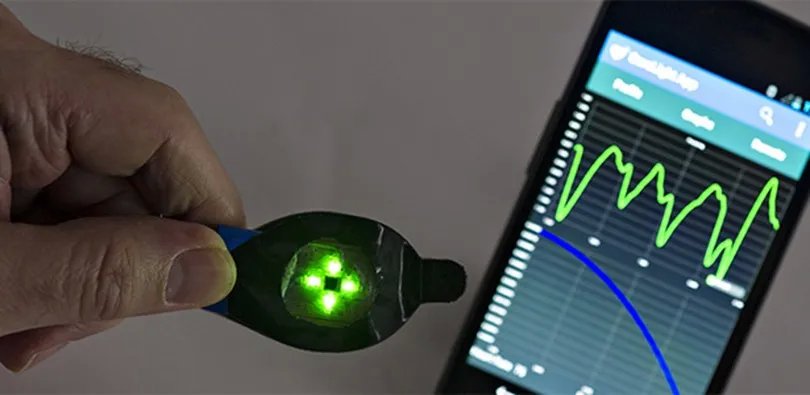
Photoplethysmography (PPG) is a non-invasive optical measurement method that detects changes in peripheral vascular bed. The word is derived from the Greek “πληθυσμóς” “plethysmos” (in- creasing), and “γρα ́φειν” “graphein” (to write) referring to the increase in blood volume in tissues due to . The basic principle of this method is relatively simple - PPG sensor is composed of a light source and a photodetector, and the PPG signal is generated by measuring the amount of light that is either absorbed or reflected by the illuminated tissue.
PPG has been studied since the mid 1930s, its wide use in clinical practice began after the ground-breaking discovery of pulse oximetry in the 1970s and its widespread among general public followed shortly after the boom of smartphones and wearables. Today PPG is the most widespread and accessible measuring modality. But interestingly, even though it has been studied for almost 100 years it is still not completely understood and the exact origin of the PPG signal is still widely debated. Research shows that the formation of the PPG signal goes way beyond the simple notion of blood volume changes in tissues and despite its simple principle, the PPG signal contains complex information about cardiovascular system reflecting rapid changes in the tissue caused by heart activity, but also slower changes caused by breathing, changes in venous filling, the influence of the autonomic nervous system and many other factors. Thorough understanding of these principles and cardiovascular pathophysiology can be the key to a next generation of low cost and widely available diagnostics and great tool for precision and predictive medicine.
Technological boom has made many measuring methods accessible to general public and of these, photoplethysmography (PPG) is probably the most widespread. Any device equipped with a light source and camera or an optical sensor such as a smartphone or a smartwatch is capable of detecting and recording changes in the microvascular bed using PPG. The idea of utilizing this technique for population-wide screening is not unique and significant financial costs and time have been already invested in pursuing of this goal. However, based on our prior and other available research we believe that we are merely scratching the surface of its possibilities. Similar to the beginnings of electrocardiography, analysing amplitudes and changes in the regularity of waveforms is only the tip of the iceberg. What at first seemed to be an endless list of patterns, originally used only for the detection of arrhythmias, now allows to diagnose many diseases that were not even known at the time of its discovery. Once a huge device weighing more than a quarter of a ton, sensitive to minimal movements, is now wearable on a belt. Equipment that once required laboratory conditions to accurately measure is now used during sports. Even the five well-known deflections P Q R S T of the ECG wave were not recorded at first but are a result of Einthoven’s mathematical correction of the original A B C D E waves.
Mathematical analysis and processing opened the door to the whole world of electrophysiology and ECG ultimately became a first-line diagnostic tool for cardiac disease.
We believe that utilising state of the art machine learning, data analysis, and our medical expertise we can take PPG diagnostics to a new level. Means to identify patients with high-risk of chronic coronary syndrome, heart failure, valvular disease or even predict deterioration of existing disease can soon be available in everyone’s pocket.
